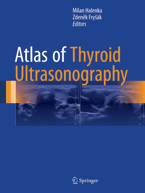
Atlas of Thyroid Ultrasonography
| Auflage | 2017 |
| Verlag | Springer |
| ISBN | 9783319537580 |
| Artikel-Nr. | 616081 |
Lieferzeit ca. 5 Werktage
Produktbeschreibung
Combining high-quality ultrasound scans with clear and concise explanatory text, this atlas includes side-by-side depictions of various conditions of the thyroid both with and without indicative marking. Each ultrasound finding is displayed twice, six figures per page: The left-hand image is a native figure without marks; the right-hand image depicts marked findings. In this way, readers have the opportunity to see the native picture to assess it by themselves and then correct their opinion, if necessary. Five sections comprise this atlas, including the normal thyroid, diffuse thyroid lesions, both benign and malicious lesions (including various carcinomas), and rare findings.
Including nearly 1500 ultrasound scans and covering the range of thyroid conditions, Atlas of Thyroid Ultrasonography will be a key reference for endocrinologists, radiologists, and primary care physicians, residents and fellows treating patients with thyroid problems.
Including nearly 1500 ultrasound scans and covering the range of thyroid conditions, Atlas of Thyroid Ultrasonography will be a key reference for endocrinologists, radiologists, and primary care physicians, residents and fellows treating patients with thyroid problems.

Bleiben Sie informiert!
Melden Sie sich für den frohberg.de-Newsletter an und nutzen Sie jetzt Ihre Vorteil:- Willkommens-Dankeschön: Beatmungsmaske Rescue Me
- Aktuelle Neuerscheinungen und Empfehlungen
- Exklusive Angebote und Kongress-Highlights
