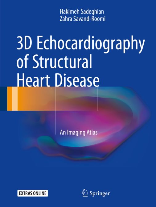
3D Echocardiography of Structural Heart Disease
An Imaging Atlas
| Auflage | 2017 |
| Verlag | Springer |
| ISBN | 9783319540382 |
| Artikel-Nr. | 559543 |
Lieferzeit ca. 5 Werktage
Produktbeschreibung
This atlas presents outstanding three-dimensional (3D) echocardiographic images of structural heart diseases, including congenital and valvular diseases and cardiac masses and tumors. The aim is to enable the reader to derive maximum diagnostic and treatment benefit from the modality through optimal image acquisition and interpretation. To this end a wide range of instructive individual cases are depicted, with sequential arrangement of all images and views of diagnostic value, including 3D zoom, full-volume, and live 3D images. For each case, a key lesson is highlighted and attention is drawn to aspects of relevance to diagnosis and treatment. In addition, readers will have online access to echocardiographic video clips for each patient. The closing part of the book examines the role of 3D echocardiography in structural heart disease interventions. The superb quality of the illustrations and the range of cases considered ensure that this atlas will be an excellent visual learning tool and an ideal aid for cardiology residents and fellows in day-to-day clinical practice.

Bleiben Sie informiert!
Melden Sie sich für den frohberg.de-Newsletter an und nutzen Sie jetzt Ihre Vorteil:- Willkommens-Dankeschön: Beatmungsmaske Rescue Me
- Aktuelle Neuerscheinungen und Empfehlungen
- Exklusive Angebote und Kongress-Highlights
