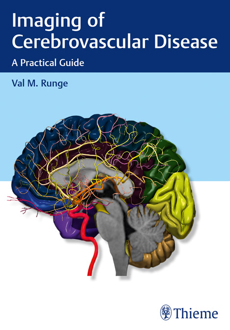
Imaging of Cerebrovascular Disease
A Practical Guide
| Auflage | 2016 |
| Seiten | 160 p., illus. |
| Verlag | Thieme |
| ISBN | 9781626232488 |
| Artikel-Nr. | 559087 |
Lieferzeit ca. 5 Werktage
Produktbeschreibung
Imaging plays an integral role in the diagnosis and intervention of debilitating, often fatal vascular diseases of the brain, such as ischemic and hemorrhagic stroke, aneurysms, and arteriovenous malformations (AVMs).
Written by a world renowned neuroradiologist and pioneer in the early adoption of magnetic resonance (MR) technology, »Imaging of Cerebrovascular Disease« is a concise yet remarkably thorough textbook that advances the reader's expertise on this subject. The text draws on the author's vast personal experience, case studies, and traditional educational sources, offering didactic dialogue with accompanying images.
A practical clinical resource organized into six chapters, this book offers unparalleled breadth in delineating the diagnostic and treatment usages of modern imaging techniques. Chapter one sets a foundation with extensive coverage of modalities and their applications, including MR, computed tomography (CT) and digital subtraction angiography (DSA). Subsequent chapters cover utilization of imaging techniques specific to underlying pathologies.
Key Features:
In-depth discussion of medical and neuroradiologic/neurosurgical interventions, focusing on the use of imaging prior to, during, and following treatment
Comprehensive text enhanced with more than 700 high-quality images
Detailed evaluation of normal brain anatomy, as well as gyral anatomy in brain ischemia, an important subtopic
Advantages, disadvantages, mortality, and morbidity of surgery (clipping) versus endovascular techniques (coiling and flow diversion) for aneurysms
Presented in a style that facilitates cover-to-cover reading, this is an essential tool for residents and fellows, and provides a robust study guide prior to sitting for relevant certification exams. It is also a quick, invaluable reference for radiologists, neurosurgeons, and neurologists in the midst of a busy clinical day.

Bleiben Sie informiert!
Melden Sie sich für den frohberg.de-Newsletter an und nutzen Sie jetzt Ihre Vorteil:- Willkommens-Dankeschön: Beatmungsmaske Rescue Me
- Aktuelle Neuerscheinungen und Empfehlungen
- Exklusive Angebote und Kongress-Highlights
