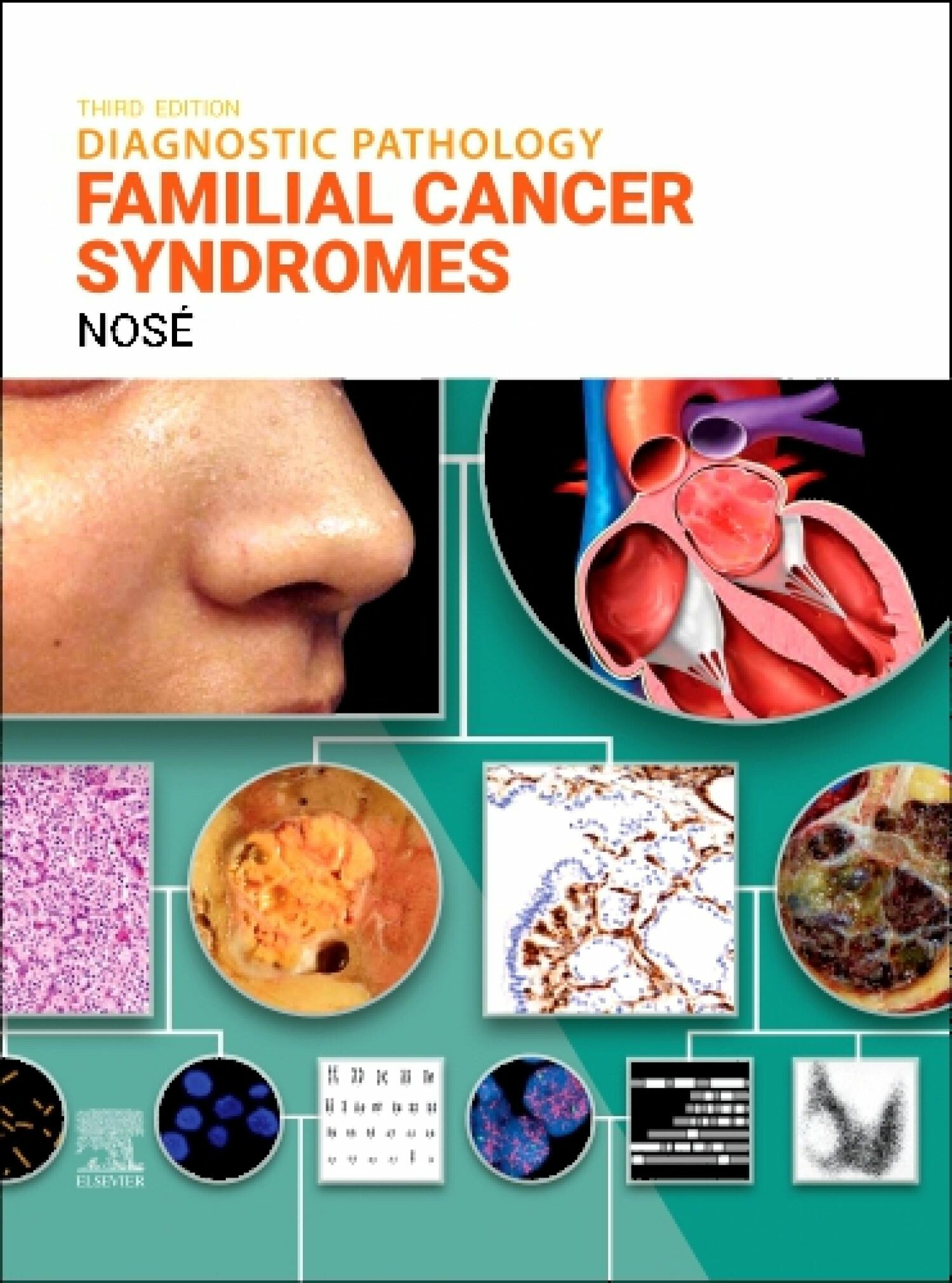
Diagnostic Pathology: Familial Cancer Syndromes - E-Book
| Auflage | 3. Auflage, 2025 |
| Verlag | Elsevier |
| ISBN | 9780443286414 |
Sofort zum Download (Download: PDF)
Weitere Formate und Ausgaben
Print-Ausgabe3rd edition 2025
€ 299,00 Print-Ausgabe
2/E 2020
€ 249,00
Produktbeschreibung
This expert volume in the Diagnostic Pathology series is an up-to-date, comprehensive diagnostic support tool for pathologists, oncologists, and other physicians who diagnose and treat patients with cancer. An excellent point-of-care reference for practitioners at all levels of experience and training, the third edition of Diagnostic Pathology: Familial Cancer Syndromes offers clinically useful information on hereditary cancer syndromes, including differential diagnosis and management. Richly illustrated and easy to use, this volume is ideal as a one-stop resource for day-to-day reference or as a reliable training tool. - Helps physicians recognize syndromes and syndrome-associated neoplasms and advise treating physicians, patients, and their families on the possibility of a familial syndrome and their risk of developing other tumors - Addresses a wide range of inherited tumor syndromes to increase understanding of the gross and histologic features of syndromic-associated neoplasms as well as associated manifestations - Part I includes nearly 100 detailed chapters describing diagnoses by organ and system associated with familial cancer syndromes; Part II contains more than 80 chapters with detailed descriptions of major inherited syndromes (cross-referenced with diagnoses); and Part III features an updated Molecular Factors Index that includes a complete description of each known gene associated with a familial cancer syndrome - Includes substantial updates throughout, with more than a dozen new chapters, new images and references, updated guidelines and classifications based on the 2025 WHO Classification of Tumours: Genetic Tumour Syndromes, details on the newest familial cancer syndromes, and more - Contains hundreds of tables organized by body part and/or organ system that include selected existing genetic and familial cancers and identified associated genes, clinical features, recognized manifestations, and staging images/microscopic examples - Features nearly 2,300 print and online images, including clinical and radiologic images, algorithms, graphics, gross pathology and histology images, and a wide range of special and immunohistochemical stains and molecular markers-all carefully annotated to highlight the most diagnostically significant factors - Employs consistently templated chapters, bulleted content, key facts, annotated images, and an extensive index for quick, expert reference at the point of care - Any additional digital ancillary content may publish up to 6 weeks following the publication date
Vania Nosé, MD, PhD, Professor of Pathology, Harvard Medical School, Pathologist and Consultant in Endocrine Pathology and Familial Syndromes, Massachusetts General Hospital, Boston, Massachusetts
Vania Nosé, MD, PhD, Professor of Pathology, Harvard Medical School, Pathologist and Consultant in Endocrine Pathology and Familial Syndromes, Massachusetts General Hospital, Boston, Massachusetts
